


")


The inspeXio 7000 is a high-performance microfocus X-ray CT system equipped with a Shimadzu microfocus X-ray generator and a large high-resolution flat panel detector.
The large detection area, input resolution equivalent to 14 megapixels, and an enhanced high-output microfocus X-ray generator enable CT images with a large field-of-view, high resolution, and high contrast. In addition, the improved HPCinspeXio high-performance computing system processes images faster.
These developments make the inspeXio 7000 system applicable for researching, developing, or inspecting a wide variety of samples, from composite materials, such as glass fiber reinforced plastic (GFRP) and continuous fiber reinforced thermoplastic laminate (CFRTP) materials to large aluminum die cast parts.
The Analytical Intelligence logo and CORE Boost are trademarks of Shimadzu Corporation or its affiliated companies in Japan and/or other countries.
VGSTUDIO MAX and VGSTUDIO are trademarks of Volume Graphics GmbH.
POLYGONALmeister is a trademark of UEL Corporation.
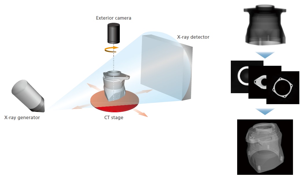
System Configuration and Operating Principle
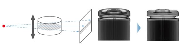
- The inspection target (sample) is placed between the X-ray generator and detector, as shown below. Then, the sample is rotated 360 degrees to collect X-ray fluoroscopic data from various angles in order to calculate cross-sectional images.
- MPR DisplayDisplays any cross section desired
Multi Planar Reconstruction (MPR) stacks multiple CT images in a virtual space to display four images—a CT image, mutually longitudinal section images, and a user-selected section image orthogonal to one of the longitudinal section images.VR DisplayVolume rendering (VR) stacks multiple CT images in a virtual space to display a 3D image. Separate 3D image processing software is required for VR display. 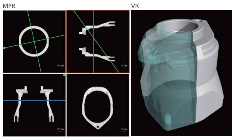
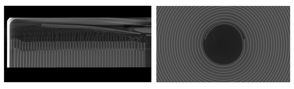
High-Resolution CT Image
Maximum 14 Megapixel Input Resolution
The large high-resolution flat panel detector achieves an offset scan input resolution of up to 14 megapixel.
Low-Resolution Cross-Sectional Image
High-Resolution Cross-Sectional Image (14 Megapixel Input Resolution)
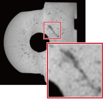
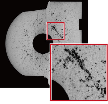
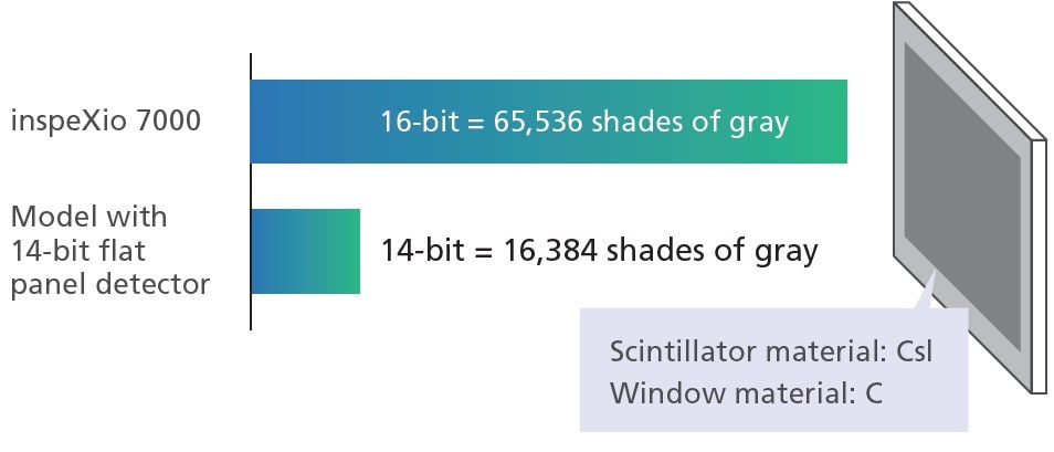
High-Contrast CT Image
High-Contrast Detector with Wide Dynamic Range
Cesium iodide (CsI), which has excellent sensitivity characteristics in the long wavelength region, is employed as the scintillator material.
The use of carbon (C) for the detector window material enables imaging on low-density materials. Furthermore, the wide dynamic range (16-bits) enables small contrast differences to be displayed.
Improved X-Ray Generator
The Shimadzu-made microfocus X-ray generator unit now includes a newly developed irradiation window. Due to the larger proportion of soft X-rays in the X-ray output, it offers significantly improved contrast when scanning low-density materials that easily transmit X-rays.
Comparison of Transmission Images from Non-Woven Fabric
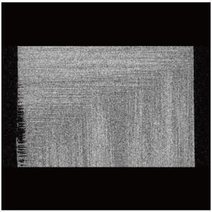
Previous Cross-Sectional Image
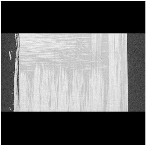
New System Cross-Sectional Image
Easy and Fast CT Scanning
Intuitive User Interface
The new user interface features a simpler arrangement for intuitive operation.
- Main System Window
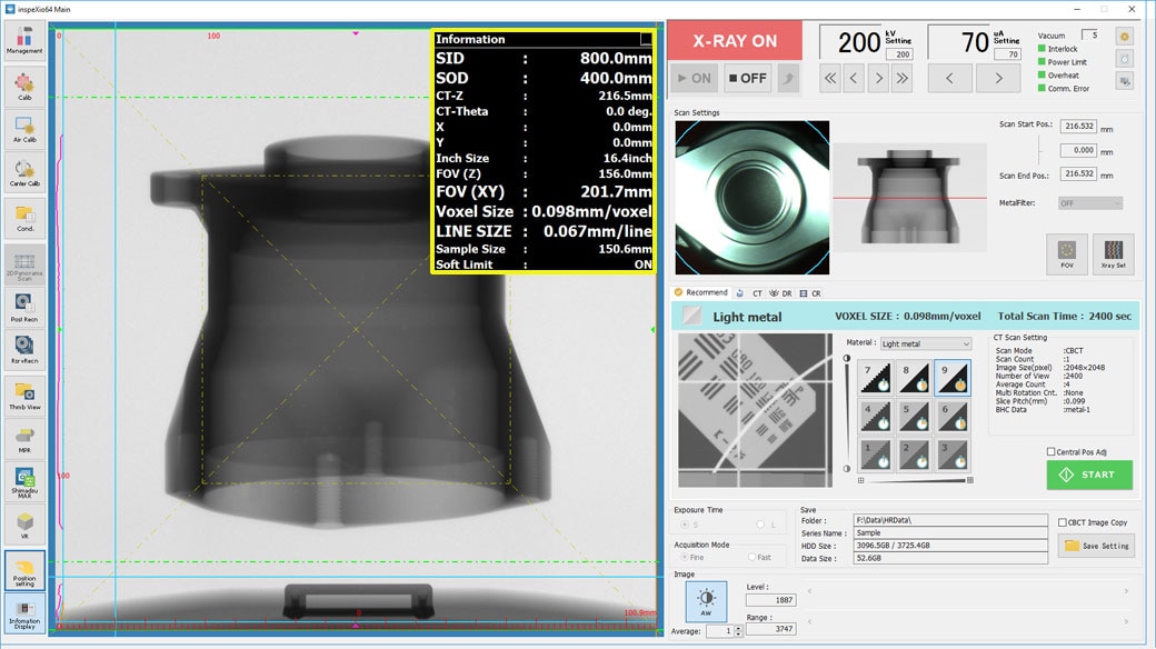 Displays the stage position, scan field of view, equivalent voxel length, and other information in real time, making (the yellow box), it easy to scan images with the specified resolution and field-of-view size.
 - MPR Window
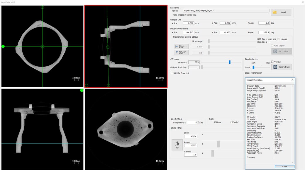 Displays slice, oblique, and double-oblique images, enabling the easy observation of cross-sections.
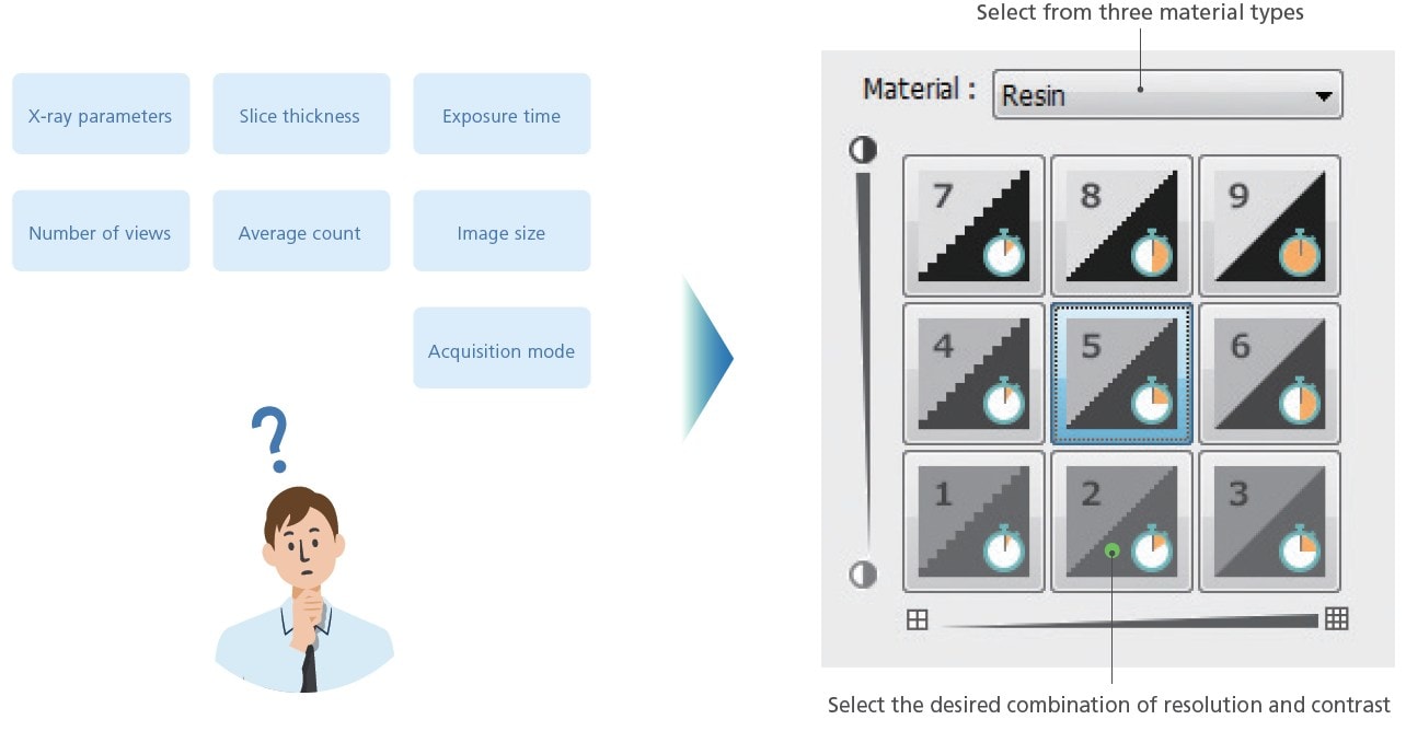
Recommend Scanning Function 
The new recommend scanning function enables scan parameters to be specified easily. Simply select the material, the desired CT image resolution, and the contrast level, and the system automatically optimizes the CT scanning parameter settings accordingly.
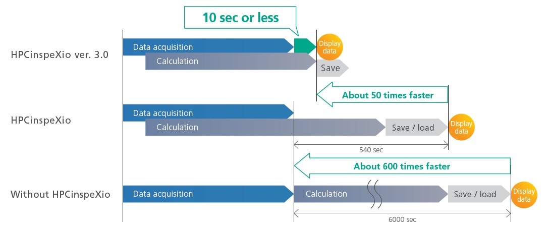
HPCinspeXio High-Performance Computing System ver. 3.0
The new HPC inspeXio high-performance computing system is around 50 times faster* than the previous version.
* When the fast acquisition mode is configured and the CT image size is set to 1,024 × 1,024 pixels
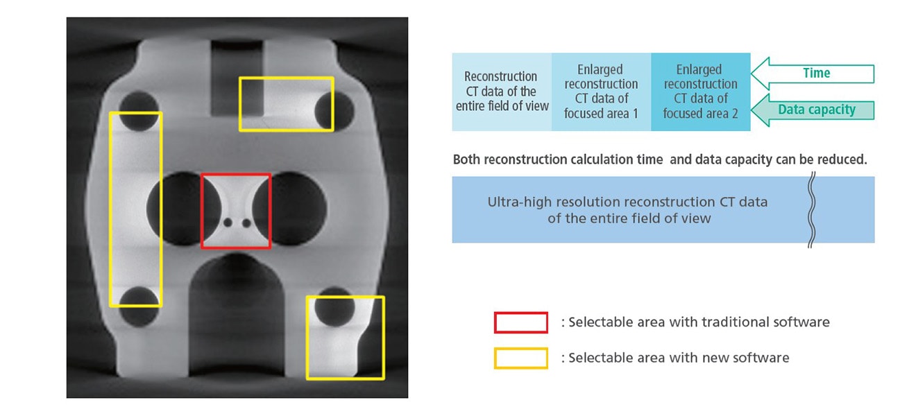
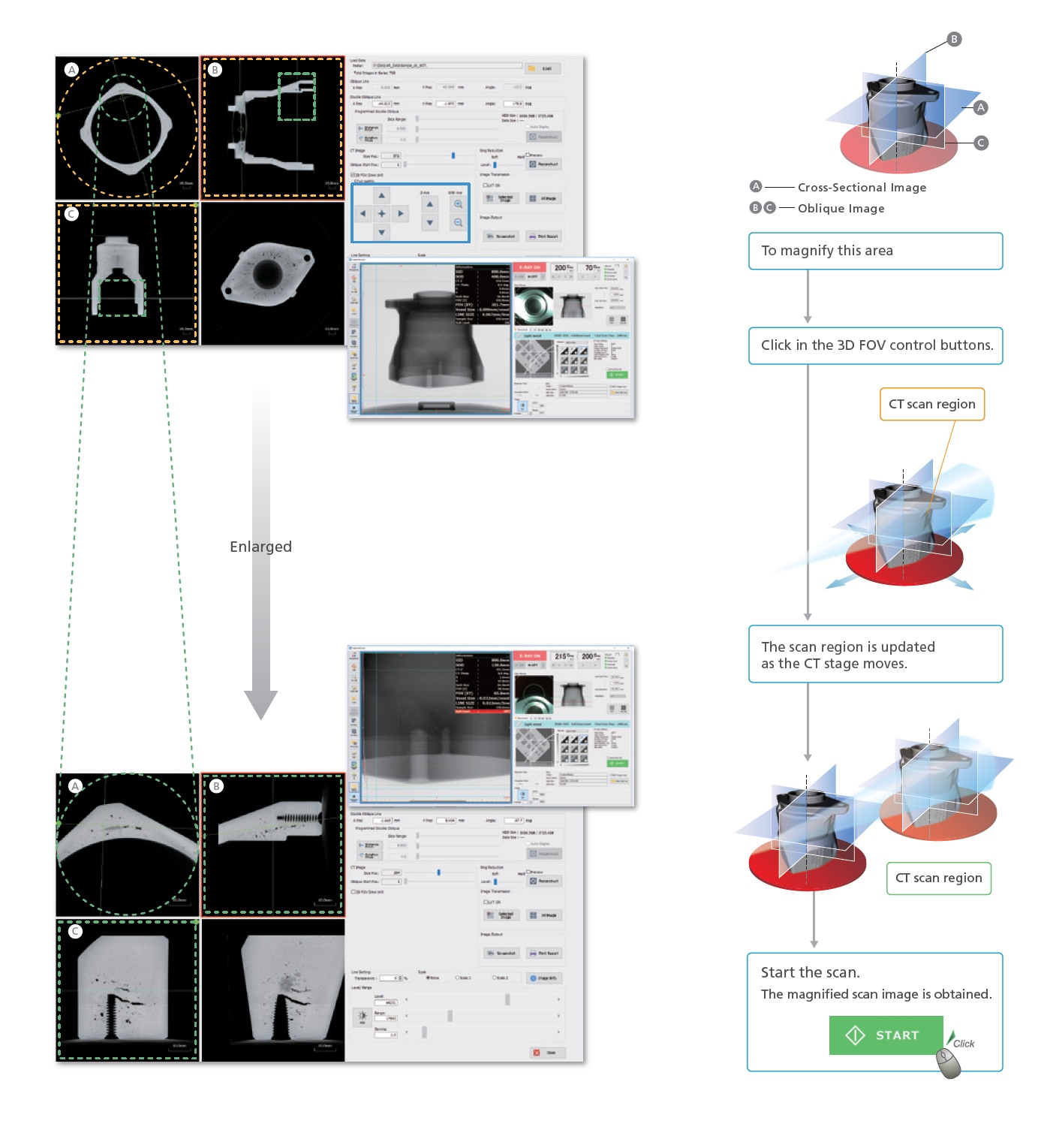
Advanced 3D image Reconstruction
It is possible to enlarge only the focused areas in images once acquired and perform the reconstruction calculation. High-magnification cross-sectional images can be obtained even in the works that enlargement ratio is difficult to be improved. Equipped with a high-resolution at panel detector, clear cross-sectional images can be obtained even when performing reconstruction. It is not necessary to perform the CT scanning once again when performing reconstruction only.
Obtain CT Images in Three Easy Steps
No calibration process is necessary before scanning. Scans can be started immediately after sample placement.
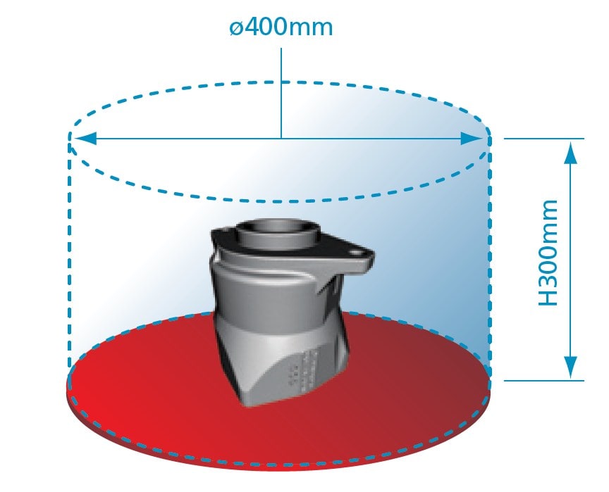
- Step1: Place the sampleMaximum sample and CT scan size are 400 mm in diameter and 300 mm in height.
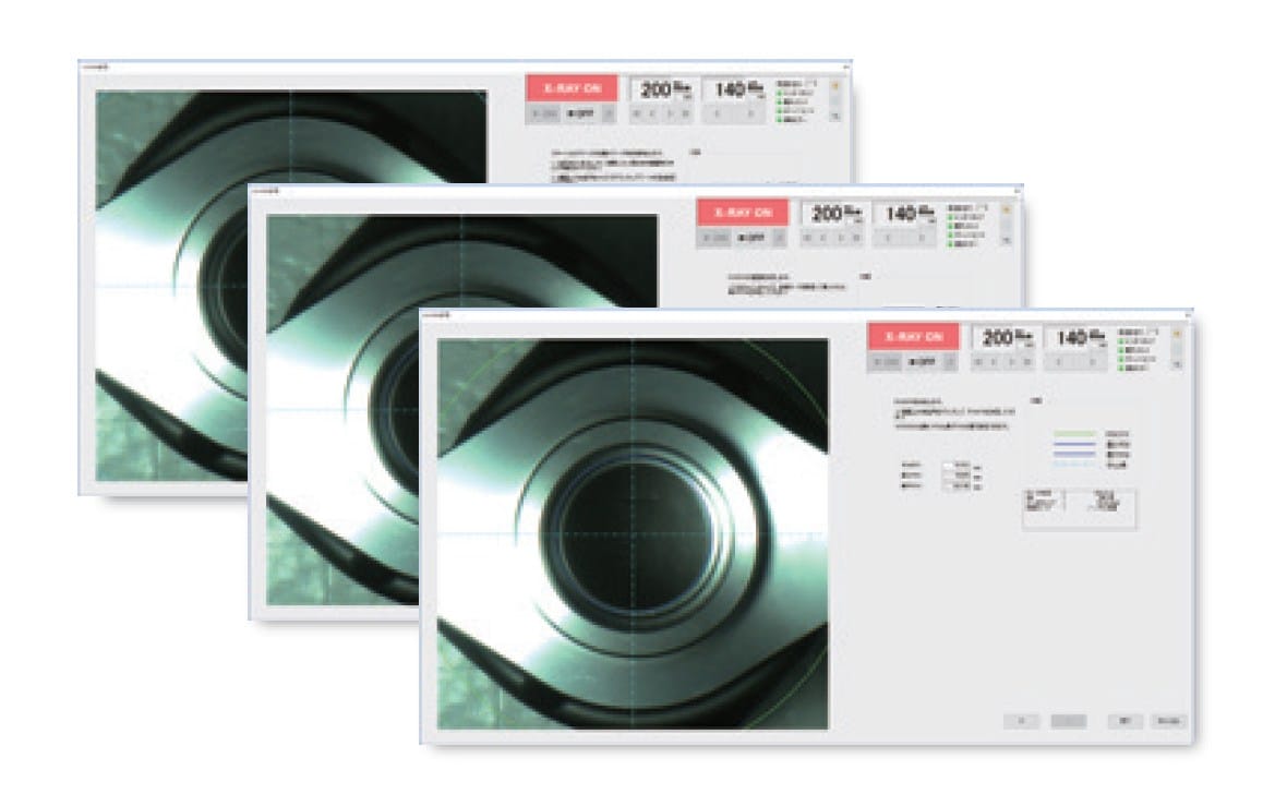
- Step2: Determine the scan positionSamples are positioned using the camera mounted on the rotation axis.
- Step3: Start the scanScans can be started immediately without prior calibration.
In normal scan (600 view), data acquisition can be done in as short as 33 seconds.
Due to the high-performance computing system, MPR images are displayed 10 seconds or less after scanning is finished. 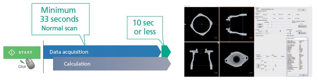
3D CT Scan Region Display Function
As the CT stage moves, the corresponding CT scan region is displayed and overlaid in real-time on the MPR display. Based on the previous CT scan image, additional CT scans for areas of interest can be obtained.
Unique Functions
- Extended Filament Lifetime
The expected lifetime of filament is extended by 2.5 times by automatically adjusting the current value. - Acquisition Mode Switching Function
Long or short scan times can be specified by combining acquisition mode and exposure time settings. - Anti-Pinch Prevention Mechanism
A finger-pinch prevention mechanism is provided to prevent accidents when closing the sliding door. - Door Interlock Mechanism
The sliding door is equipped with redundant interlock circuits. These ensure X-rays are never emitted when the sliding door is open. In addition, these stop the CT stage from moving when the sliding door is open. - CR Scan
- Computed radiography (CR) can be used to obtain transmission images without distortion in the CT-Z direction by acquiring data only along the vertical center line of the X-ray detector while moving the CT-Z axis vertically.
- DICOM Conversion Function
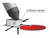
CT Image data can be converted to the DICOM format, which is the world standard for medical imaging. Consequently, this function is essential for analyzing data with medical image analysis software. - Collision Sensor
- The expected lifetime of filament is extended by 2.5 times by automatically adjusting the current value.
Improvement of S/N Ratio by SID Switching Function
Source to Image Distance (SID) can be switched among three settings. Depending on the size and area of the object to be imaged, the flat panel detector can be positioned closer to the X-ray generator to obtain a stronger signal and thereby reduce data noise.
4680-type lithium-ion rechargeable battery

